Osteodystrophia deformans: Paget’s disease of bone
In the case of chronic disease of the bone metabolism, Paget’s disease (Osteodystrophia deformans) is a pathologically increased bone remodeling, leading to thickened, deformed and brittle bones. Many of those Affected remain free for a long time symptom, but it is also drilling bone pain and other discomfort and serious consequence can occur. Often a diagnosis is made relatively late, which may affect the success of treatment. The therapy for Paget’s disease includes, in addition to analgesic drugs, especially the administration of newer bisphosphonates, to inhibit bone resorption.
Important note: The term Paget’s disease is used interchangeably for two different clinical pictures. The first one is a disease of the female breast (Paget’s carcinoma) in the other, a bone disease, which is also known as Osteodystrophia deformans or Osteitis deformans. The following article includes exclusively the bone disease.
Table of contents
A brief Overview
In addition to the below comprehensive information on the bone disease, Paget’s disease, summarizes the following is a brief Overview of the main facts of this relatively rarely diagnosed disease:
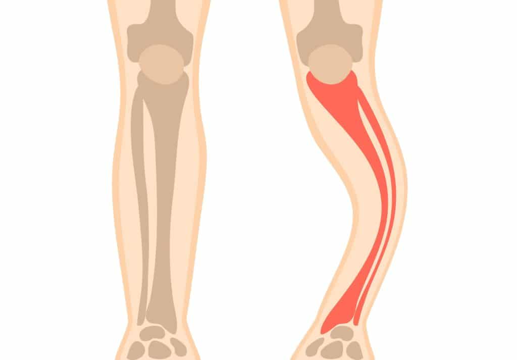
- Definition: Osteodystrophia deformans, is a chronic disease of the skeletal system, in which a disturbed bone metabolism process. A pathologically increased bone remodeling leads to a gradual thickening and deformation of the affected bone, which also have a lower load capacity and stability.
- Symptoms: The disease develops in about 90 percent of the Patients in the long term, symptom-free. It complaints felt, pain is the first Symptoms are local, and in-depth bones, and you may also have other pain and discomfort. The deformations can be visible and the pathological processes are partly in the Form of heat through the skin noticeably.
- Causes: The causes are as yet unclear. It is a hereditary components are discussed in connection with a unknown virus infection as a trigger.
- Diagnosis: A diagnosis is often late, or even not possible. The presumption on Paget’s disease, serve in the first line of an x-ray examination, bone scintigraphy and a blood test to further diagnosis.
- Treatment: Symptom relief and inhibition of bone remodelling are mainly drug therapies with bisphosphonates and Calcitoninen. Under certain circumstances, can also painkillers or, in severe cases, surgery can be considered. In General, the treatment is complemented with the intake of Vitamin D and calcium, as well as with physiotherapy.
Definition
The name Osteodystrophia deformans (Osteitis deformans) means “deformation of the bone”, which is the time the disease is already roughly described. The synonym terms used Paget’s disease, Paget’s disease or Paget’s disease relate to the first descriptive surgeons and pathologists by the name of James Paget of England, where the disease actually occurs most frequently. The first describer referred to in the 19th century. A century on, an increasing hat size as a first sign.
Today, we define Paget’s disease as a chronic disease of the skeletal system, which leads to a disturbed bone metabolism process, and in one or more areas of performance. The pathological changes involve a disorganized bone remodeling and a gradual thickening and deformation of the affected bone. In addition, there is a lower load-bearing capacity and stability of the modified bone structure.
The disease occurs, as a rule, only in the case of older people, especially after the age of 40. or after the age of 55. Years of age. The majority of disease cases are to be found in Northern Europe (especially England and, with the exception of Scandinavia). In Asia and Africa, the Occurrence of Osteodystrophia deformans is, however, very rare. Overall, more men are affected than women. In General, the disease is one of the rare Osteopathien (sick bone changes). However, a high number of unreported cases is suspected.
Symptoms and possible sequelae
Approximately 90 percent of the Paget’s disease Patients long-term symptom-free course of disease is observed. In the case of the remaining ten percent of the complaints of a gradually progressive developing process.
The first main symptoms are local bone pain that are often felt as a deep pain. Sometimes, the first, inflammatory processes and abnormal metabolic processes are increased through the skin in the Form of heat significantly (Hyperthermia). Affected are mostly heavily loaded bones such as the pelvis, the leg bones, spine, upper arm bone, and partly also the jaw bone. But also on the skull bone, the disease often manifests itself.
The altered and fragile bone structure leading to (visible) deformations, painful bone damage, and possibly also to bone fractures. As a further consequence of arthritis forming, leading to joint pain. The deformation may be accompanied by stress, can also cause muscle pain, caused by muscle spasms or Muscle tightness.
It is due to the distention of the bone to an excessive pressure on nerves, may be affected in severe cases, also their functions. As a secondary complaints can pain, for example, in the area of the spine, Sciatic, back pain and low back pain occur. In the case of an infestation of the skull, various factors can cause, such as Sensorineural or conduction disturbances, even a hard of hearing (hypoacusis).
A rare (less than one percent) complication is the emergence of a malignant tumor (bone cancer). In these few cases, it is usually a secondary osteosarcoma. Also very rarely it comes to heart overload by increased blood flow.
At about ten to fifteen percent of Patients the elevated calcium required for the increased bone formation to the formation of a secondary hyperparathyroidism (Hyperparathyroidism) and hypocalcemia (calcium deficiency) and hypercalcemia (calcium excess). In the Presence of hyperparathyroidism, which is formed parathyroid hormone (parathyroid hormone) to regulate the calcium level in the blood.

Causes
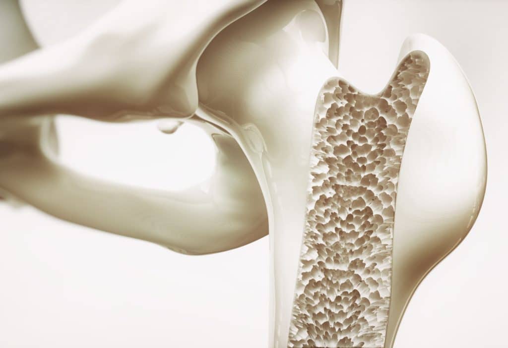
The emergence of the disease is still unclear. So far, it managed to subject-matter experts to identify several genetic mutations associated with the development and activity of the so-called osteoclasts. The often large and multinucleated osteoclasts cause Resorption of bone tissue and to show the Paget’s disease Patients, increased activity. The capacity of the osteoclasts can be a multiple of the so-called osteoblasts, which are, in turn, for the formation of bone tissue responsible.
Furthermore, the affected bone can exact considerations a viral infection suspect. Thus, it is assumed that at a certain genetic predisposition, an unknown Virus triggers the abnormal osteoclast activity, which in turn caused the specific damage to your Bones. A proof for this theory has not been provided yet.
Diagnosis
Deformed limb to fall already (usually asymmetrical), comes relatively quickly, the suspicion of Osteodystrophia deformans. However, since in many patients cases, over long periods of time may not experience any discomfort or are visible and a characteristic clinical picture is missing, late or not at all, or Paget, an assumption and a diagnosis of Crohn’s it is often the case.
A suspected Paget’s disease can be made to diagnose, as a rule, the first x-ray images. This imaging method allows for relatively early detection of bone degradation (osteolysis). Also findings such as deformation or thickening of the affected bone or osteoporosis can be created by means of x-ray images. The bone scintigraphy is another imaging method of the nuclear medical diagnosis, which is in addition an increased bone remodeling can be made visible.

Usually is performed for further Diagnosis, a blood test, in which certain enzymes (alkaline phosphatases) in the Serum can be determined. In the majority of Paget’s disease and non-Diseased, are still in therapy, for these enzymes, elevated values. In addition, other markers can indicate increased bone resorption, such as, for example, the so-called C-terminal Telopeptide (CTX) in the Serum or released amino acids (hydroxyproline) in the urine.
A bone biopsy can, in cases of doubt, or in the case of a complicated course of illness with suspected osteosarcoma. So also in the case of certain complications, for further investigation techniques such as computed tomography and magnetic resonance imaging may be indicated.
Treatment
For a long time, the disease was considered to be not or only partially treatable. Nowadays, personalized and, above all, at an early stage, begun in therapies is attributed to a relatively high success rate, in order to delay the progression of the disease. Existing deficits are often irreversible.
The goal of any treatment is to relieve the pain and symptoms and to reduce, if possible, also the bone loss and the threat of complications. The individual characteristics determine the priorities of a therapy concept. So is usually the first goal is to reduce the bone deformations when mechanically highly loaded bones are affected (for example, the pelvis or the legs). However, it may be a infection of the spine or the skull, a first target, to prevent possible nerve damage.
In the treatment is generally drug therapy, especially with the use of bisphosphonates and Calcitoninen. These drugs are intended to inhibit the osteoclast activity and the resulting bone resorption. The first choice is usually a bisphosphonate, such as Etidronate, Pamidronate, or Zolendronat. It comes to intolerance of or resistance to a bisphosphonate, is often made of synthetic salmon Calcitonin. The newer bisphosphonates (for example, Zolendronat) are usually more effective and longer effective.

The importance of a timely and regular intake of medicines is, in any case. Some bisphosphonates can be administered for a long-term effect (up to one year), also intravenously. A follow-up of this therapy is usually, as mentioned in the diagnosis, with the help of a blood test carried out. By the determination of the alkaline phosphatases, the activity of the osteoclasts can be estimated in the course of the disease.
In the case of existing pain, usually in addition to pain-relieving and anti-inflammatory medications are used such as non-steroidal anti-inflammatory Drugs, or similar drugs.
Many of those Affected can help in addition, targeted physiotherapy, resulting stress, to counteract and to maintain the General mobility. For further Symptom relief, particularly in older Affected, other treatments such as a Massage or electro-therapy.
The physical impairments can be so strong that the possibility of surgery is considered. Possible surgical procedures, the osteotomy, the bone is severed and re-fixed to be, or a bone or joint replacement by endoprosthesis.
Due to the increased calcium requirements for the accelerated bone remodeling and may result in calcium deficiency, will advise the Affected often have a supplementary intake of calcium and Vitamin D. Vitamin D (or the prohormone Cholecalciferol) acts on the calcium metabolism and strengthens bones.