Researchers at UC have discovered a previously unknown mechanism that could explain the reason behind decreased immune function in cancer patients and could be a new therapeutic target for immunotherapy for those with head and neck cancers.
The authors share these findings in an article published in the journal Frontiers in Pharmacology.
Immunotherapy is a type of cancer treatment that boosts the body’s natural defenses to fight cancer.
Led by Laura Conforti, professor in the Department of Nephrology and Hypertension at the UC College of Medicine, the team discovered that a reduced interaction between a molecule called calmodulin and an ion channel (KCa3.1) in the immune cells of cancer patients plays an important role in the reduced function of these cells. The team performed experiments on white blood cells called cytotoxic T-cells taken from the blood of patients with head and neck cancer.
“Cytotoxic T-cells are like the soldiers of our immune system and are our body’s first line of defense against cancerous tumors,” says first author Ameet Chimote, research scientist in Conforti’s laboratory. “Just like how soldiers on the frontlines penetrate the enemy’s defense and launch a massive attack, these cytotoxic T-cells are expected to penetrate the solid tumors by migrating within the tumor mass and then secreting chemicals called cytokines to kill these tumor cells. Sadly, for some reason, these cells do not function properly in patients with cancer, and they do not penetrate the tumors and attack the tumor cells, causing the cancerous tumors to grow uncontrollably.”
“Identifying the mechanism of this underlying dysfunction can help us identify molecules that we can target with drugs and ultimately restore the ability of these cells to enter and kill the tumors,” says Conforti. She says that molecules, known as ion channels, are present in the T-cell membranes and are essential for T-cell function.
“In this study, we were able to show that the function of these channels in T-cells from cancer patients is decreased which results in a decreased T-cell accumulation in solid tumors,” Conforti says.
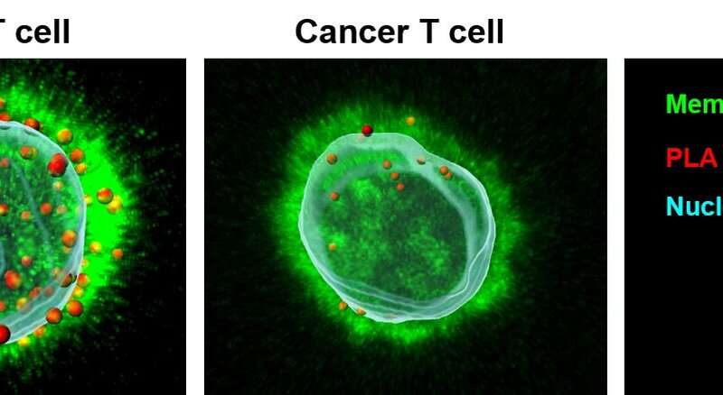
These types of channels require a signaling molecule called calmodulin to bind to them in order to function to their full capacity; this is needed even more in cancer T-cells.
Using several intricate microscopy imaging techniques on T-cells isolated from the blood of cancer patients, the team found out that, as compared to T-cells from healthy individuals, the cancer T-cells have fewer calmodulin molecules in their membranes.
“This would mean that there is less calmodulin binding to the channels in the T-cells from cancer patients,” Conforti says. “As previously stated, the channels do not function if the calmodulin does not bind to them. Thus, the decreased calmodulin binding in T-cells from cancer patients results in decreased function and leads to reduced tumor infiltration and killing of the cancer cells.”
“So back to our soldiers: If they were present at a battlefield, but none of them have any weaponry, this will hinder their ability to infiltrate the enemy lines. Now, if we arm each soldier adequately, we would boost their function. They can do their job. Similarly, we observed that if we increase the function of these channels by drugs that enhance their activity, the cancer patients’ T-cells can penetrate the tumors better and also produce increased cytokines which can kill tumor cells,” Chimote says. “These are exciting findings that could lead to additional treatments for patients with cancer.”
Source: Read Full Article
