 insights from industryDr. Jason OtterstromApplication ScientistIDEA Bio-Medical
In this interview, NewsMedical talks to Jason Otterstrom about the role of AI-based Athena Zebrafish software in exploring Zebrafish imaging results and its importance for bio-medical research using Zebrafish models. Athena is now available for download with a flexible pay-per-use license.
How can high-content imaging of Zebrafish revolutionize the bio-medical research field?
Zebrafish (Danio rerio) are a promising emergent model system for biomedical research with the potential to enable fast, high throughput studies of human disease in living vertebrates.
Humans and zebrafish have most of the same organs, share 70% of the same genes, and 84% of the genes associated with human disease have zebrafish counterparts.
Zebrafish embryos are transparent and can be manipulated at the single-cell stage, then quickly grow into larvae in a matter of days. This physiological development can be imaged with a microscope using thousands of individual fish to directly visualize the effect of treatments on organ systems, development and physiology.
No other well-characterized biological model system is currently able to offer these advantages.
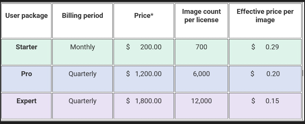
Dr. Otterstrom: "We offer three affordable license packages for different throughput needs. With an effective price between $0.15 and $0.29 USD per image analyzed, any Zebrafish lab or researcher can use Athena software to enhance their analysis workflows." Image Credit: IDEA Bio-Medical Ltd.
How are images of zebrafish used, and why are they important for researchers?
Microscopy images of zebrafish are analyzed to extract numerical or quantitative information about the embryos and larvae. The animal's transparent body makes much of the fish’s internal anatomy observable, even with simple bright-field imaging.
Genetic manipulation permits the insertion of fluorescent proteins into the live zebrafish for tissue-specific imaging and analysis.
Life science researchers then analyze images of zebrafish to study and measure biological and physiological changes happening in different anatomical regions within a living fish. For example, organ development and cellular proliferation or death in relation to pharmacokinetics, toxicity, cancer, and genetic changes can be directly observed and measured.
How can AI help in these investigations?
Analyzing images of zebrafish is a time-consuming bottleneck in screening studies. While the images may be easily interpretable by a human, conventional image analysis algorithms do not readily detect the fish or its anatomy. Instead, measurements are performed manually using single images.
IDEA Bio-Medical learned of this challenge from our clients, so we created a novel deep-learning AI (artificial intelligence) algorithm to automatically analyze their Zebrafish images for them.
The AI training process is complex and error-prone, requiring substantial time and effort that our biology-focused clients could not do themselves. The AI we have developed can now robustly identify objects like the zebrafish contour and its internal anatomy that are clearly visible to us humans.
We now want to make this tool available to all zebrafish researchers, no matter which microscope they use.
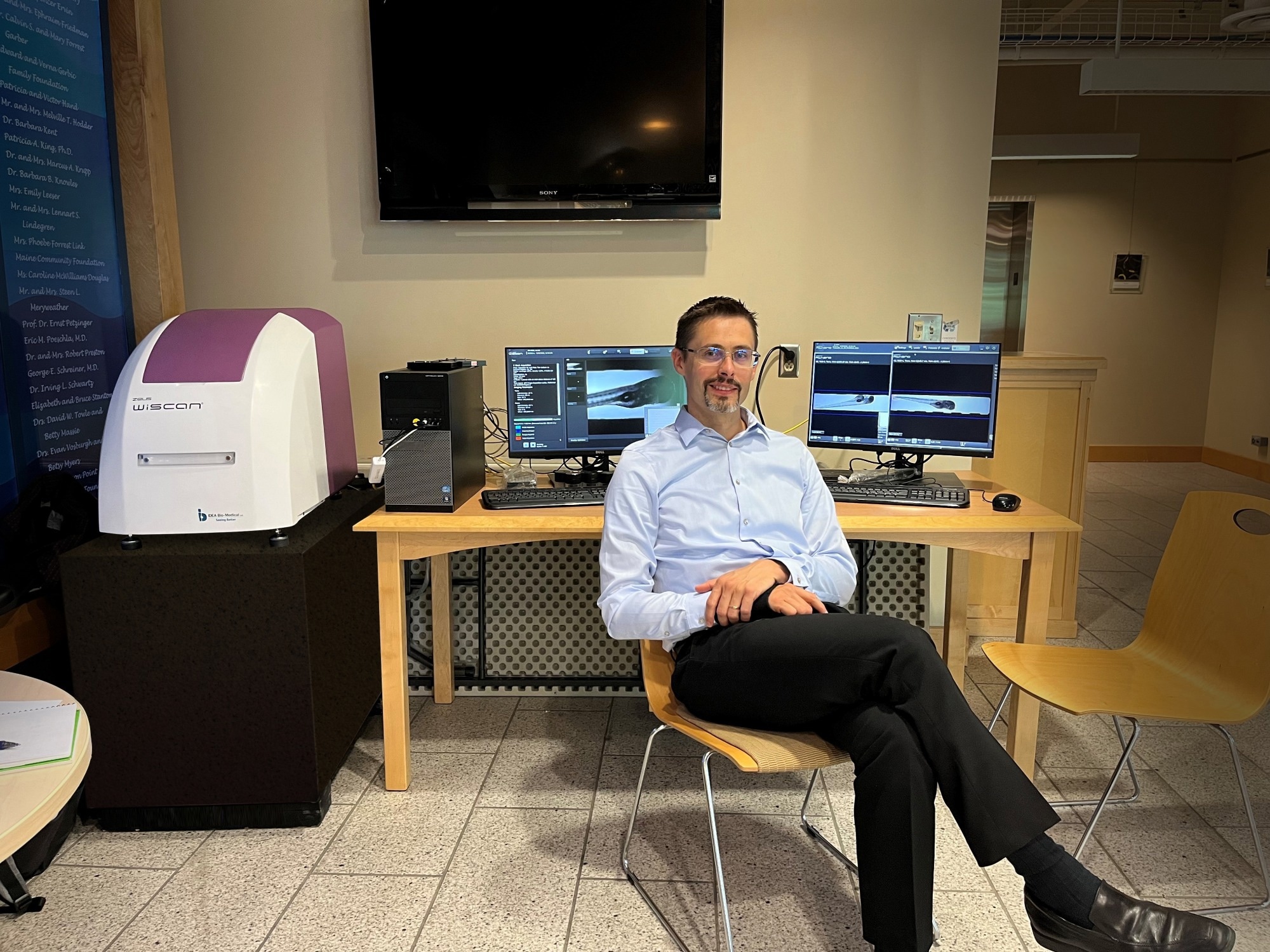
Athena software permits parameter-free Zebrafish analysis using simple bright-field images, extracting the fish contour and much of its internal anatomy (yolk sac, eye, notocord and more), along with body regions of the head, trunk, and tail. Image Credit: IDEA Bio-Medical Ltd.
Why do we need information about Zebrafish anatomy to interpret fluorescence signals?
Many processes investigated using zebrafish take place or begin in specific organs or regions within the body. They can be marked with fluorescence labeling to image the tissue or cells of interest. However, these must be distinguished from undesired signals and autofluorescence.
For example, in the zebrafish larva, hematopoietic stem cells (HSC) originate in a specific area within the tail – otherwise known as the caudal hematopoietic tissue (CHT). It is analogous to the mammalian fetal liver and relevant to leukemia in humans. Studies looking at the proliferation of GFP-labeled HSC need to focus specifically on the tail and ignore those fluorescent signals present elsewhere, such as circulating cells and yolk sac autofluorescence.
Athena is currently the only software available that enables high-content screening in this context. It permits automated cell counts exclusively within specific parts of the fish to maximize both throughput and data extracted.
It contrasts common approaches, such as simple intensity thresholding, which ignores the visible anatomy. Instead, all fluorescence is gathered together, irrespective of its origin, producing noisy data. Manual selection of the tail would improve quality but severely limit achievable throughput.
Athena is a first-of-its-kind automated image analysis software dedicated to zebrafish microscopy. Image Credit: IDEA Bio-Medical Ltd.
What is Athena image analysis software, and how can it be used?
Athena Zebrafish is a stand-alone software developed and offered by IDEA Bio-Medical to empower all zebrafish researchers. It enables true high-content imaging in zebrafish for the first time.
Athena permits parameter-free zebrafish analysis using simple bright-field images. It automatically detects zebrafish embryos and larvae up to 5 days old (dpf), extracting the fish contour and much of its internal anatomy (yolk sac, eye, notocord, and more), along with body regions of the head, trunk, and tail.
For each of these objects, the software measures the morphology (area, length, and shape) and can detect fluorescence in associated color channels. Both fluorescence intensity and spot/structure detection within specific anatomy are supported.
The software is suited for a broad range of researchers and accepts multiple image format types output from nearly all microscope manufacturers.
Click here for a free 14-day trial of Athena Zebrafish software, available for download from the IDEA Bio-Medical website.
What advantages does this method have over alternative techniques?
Athena is the only automated software dedicated to zebrafish microscopy, offering several advantages:
- Automation: It allows experimental scale-up while eliminating subjective human bias or error and permitting direct comparison of results between different labs.
- Intuitive and user-friendly interface: Users can quickly and easily adapt Athena for their research.
- Versatile and flexible AI: Widely applicable for many types of screens involving zebrafish, as described by Athena customers on the IDEA Bio-Medical YouTube page.
- Customer support with personalized new user onboarding to ensure the success of our customers.
What is the Flexible Pay-Per-Use license you are offering, and what are its advantages?
IDEA Bio-Medical recognizes that annual software licenses can be expensive, especially if images are not consistently analyzed throughout the year. Hence, we offer Athena on a pay-per-use basis so that zebrafish researchers can adapt their license to analyze images when they need it.
We offer three affordable packages for different throughput needs. With an effective price between $0.15 and $0.29 USD per image, any Zebrafish lab or researcher can use Athena and enhance their analysis workflows.
Licenses can be purchased monthly or quarterly, according to need. This flexibility allows labs to adjust their licenses to fit their changing projects, students, grant submissions and other dynamics of academic research.
Source: IDEA Bio-Medical Ltd.
What support does IDEA Bio-Medical provide to Athena software users?
Analyzing microscopy images is complex, requiring skill and experience. Along with an intuitive software interface, we offer personalized customer support to enable any user of any skill level to become independent and productive with Athena.
We at IDEA Bio-Medical take pride in working closely with our clients and their unique needs. New users can receive a one-on-one onboarding session with a qualified product specialist to walk through the software using their own data and ensure a smooth, positive first experience.
Additional Athena support is also available by reaching out to IDEA Bio-Medical directly.
What is the future direction of your Athena software, and who would benefit from using it?
Athena is a rich application-based image analysis software for easy and quick analysis of microscope images. The full Athena package is currently integrated into the Hermes platform, IDEA Bio-Medical’s flagship high content screening (HCS) microscope.
In the future, the full Athena software, relevant for microscopy images of 2D and 3D cell culture, will be offered as a stand-alone platform, allowing all biology researchers to perform image analysis at the push of a button.
About Dr. Otterstrom
Dr. Otterstrom obtained a Bachelor of Arts in Applied Physics at the University of Utah. As a Ph.D. student at Harvard University, he studied the biophysics of membrane fusion as mediated by influenza virus hemagglutinin protein using single-molecule microscopy techniques. He went on to obtain a Marie Curie fellowship to utilize super-resolution imaging to study chromatin fine-packing structures at the Institute of Photonic Sciences (ICFO) near Barcelona, Spain.
As an application scientist for IDEA Bio-Medical, he supports clients with adapting their diverse experiments and assays to be performed in the context of automated microscopy on the company’s flagship product, the WiScan Hermes.
About IDEA Bio-Medical Ltd.
IDEA Bio-Medical was founded in 2007 through a partnership between YEDA (the Weizmann Institute’s commercialization arm) and IDEA Machine Development (an innovation hub).
The company specializes in automated imaging systems and image analysis software, offering a broad range of biological applications based on the company’s unique algorithms library.
The WiScan Hermes system incorporates the most advanced technologies currently available in the machine vision field, integrated with engineering methodologies of high reliability and quality at the level of semi-conductors and digital printing industries, which are the specialty of the mother company, IDEA Machine Development Design and Production Ltd.
Sponsored Content Policy: News-Medical.net publishes articles and related content that may be derived from sources where we have existing commercial relationships, provided such content adds value to the core editorial ethos of News-Medical.Net which is to educate and inform site visitors interested in medical research, science, medical devices and treatments.