SARS-CoV-2 Eris variant spreads faster and dodges immunity

In a recent study posted to the bioRxiv preprint* server, researchers in Japan investigated the proliferative capacity, transmissibility, and pathogenicity of the severe acute respiratory syndrome coronavirus 2 (SARS-CoV-2) EG.5.1 variant, dubbed "Eris."
EG.5.1, an isolate derived from the severe acute respiratory syndrome coronavirus 2 Omicron variant of concern's XBB subvariant, has rapidly increased in prevalence globally. Compared to XBB.1.5, the EG.5.1 variant has additional mutations within its spike (S) glycoprotein, i.e., F456L and Q52H. However, data on the transmissibility, pathogenicity, and immune evasiveness of the EG.5.1 variant are limited.
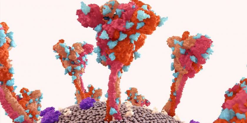
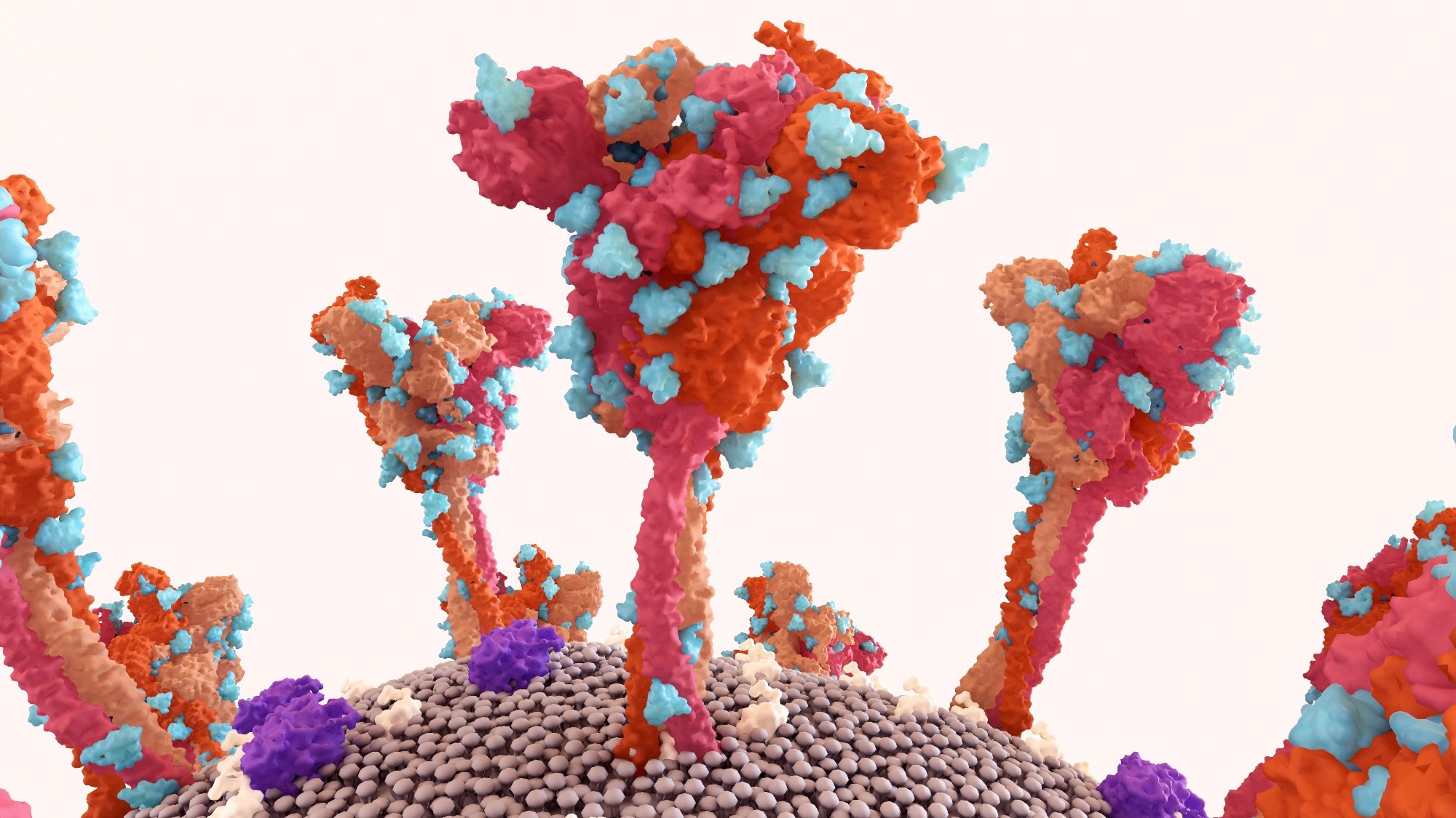
 Study: Characterization of an EG.5.1 clinical isolate in vitro and in vivo. Image Credit: Design_Cells / Shutterstock

 *Important notice: bioRxiv publishes preliminary scientific reports that are not peer-reviewed and, therefore, should not be regarded as conclusive, guide clinical practice/health-related behavior, or treated as established information.
About the study
In the present study, researchers performed virological characterization of EG.5.1 in vitro as well as in vivo.
The team evaluated the antigenicity, transmissibility, antigenicity, proliferative capacity, and pathogenicity of the EG.5.1 variant in a wild-type Syrian hamster model that is susceptible to SARS-CoV-2. In addition, they assessed the growth ability of the viruses in respiratory organs. EG.5.1, containing two additional amino acid changes (i.e., F456L and Q52H), compared to an XBB.1.5 isolate, was amplified.
To evaluate EG.5.1 immune-evasiveness, the team tested the EG.5.1 neutralization potential of serum obtained from messenger ribonucleic acid (mRNA) vaccine recipients who experienced breakthrough infections with SARS-CoV-2 variants in circulation post-March 2023. To evaluate the effects of the EG.5.1 variant mutations, the XBB.1.5 and XBB.1.9.2 strains of SARS-CoV-2 were used for comparative assessment.
African green monkey (Vero E6) cells expressing angiotensin-converting enzyme 2 (ACE2) and transmembrane protease serine 2 (TMPRSS2) enzymes were used for the cell culture experiments and confirmed to be mycoplasma-free by polymerase chain reaction (PCR). The neutralization capacity of human serum was expressed as 50% focus reduction neutralization titers (FRNT50).
For pathological and virological assessments, wild-type Syrian hamsters were intranasally inoculated with 105 plaque-forming units (PFU) of SARS-CoV-2 Delta, EG.5.1, or XBB.1.5 variants. At three and six days of infection, five animals were sacrificed, and samples of their nasal turbinates and lungs were examined for histopathological changes. Plaque assays were used to determine viral titers, and viral RNA, extracted from the tissues, was subjected to whole genome sequencing (WGS) analysis.
Results
The team found no significant differences in pathogenicity and growth ability between XBB.1.5 and EG.5.1, and both strains were attenuated in comparison to the Delta variant. In addition, EG.5.1 transmitted more efficiently among hamsters than XBB.1.5. Unlike XBB.1.5, EG.5.1 was detected in the lungs of four out of six infected hamsters, indicating that EG.5.1 virus tropism differed from that of XBB.1.5 following transmission through air.
EG.5.1 neutralization was marginally, but significantly, lower than XBB.1.5 or XBB.1.9.2 neutralization, indicating that EG.5.1 effectively evaded antibody-mediated immunity and that the amino acid differences in EG.5.1 spike (S) glycoprotein compared with that of XBB.1.5 or XBB.1.9.2 (i.e., R158G, F456L, and Q52H) altered EG.5.1 antigenicity. The Delta infection caused considerable weight loss in the animals. In contrast, all XBB.1.5- and EG.5.1-infected animals gained body weight over 10 days, to a similar extent as the mock hamsters.
After three and six days of infection, the XBB.1.5 variant and the EG.5.1 variant proliferated in the pulmonary tissues of the SARS-CoV-2-exposed hamsters with no statistically significant differences. Nevertheless, the viral titers within the nasal turbinate specimens obtained from the SARS-CoV-2 EG.5.1-exposed animals were significantly greater compared to those among XBB.1.5-exposed animals at six dpi. Additionally, the viral titers in Delta-infected hamsters were significantly higher than those among XBB.1.5- and EG.5.1-infected hamsters.
The team observed marginally greater efficacy of transmission of the EG.5.1 variant (83%) compared to that of the XBB.1.5 variant (56%) and viral detection in the pulmonary tissues of 67% of EG.5.1-infected hamsters, contrasting no virus identified in those of their XBB.1.5-infected counterparts. Although all serological specimens showed neutralizing activity against the EG.5.1variant, the FRNT50 titers against the EG.5.1 variant, the XBB.1.9.2 variant, and the XBB.1.5 variant were lower by 10-fold, 5.4-fold, and 5.5-fold compared to the titers against the SARS-CoV-2 Wuhan-Hu-1 strain, respectively.
Conclusions
Overall, the study findings showed that EG.5.1 had a similar proliferative ability to XBB.1.5 in naïve wild-type hamsters; however, its transmissibility was somewhat greater. Furthermore, the viral tropism of the EG.5.1 variant differed from the viral tropism of the XBB.1.5 variant following transmission via air since the EG.5.1 variant was found in specimens obtained from the nasal and pulmonary tissues of infected hamsters. EG.5.1 immune evasiveness was modestly but considerably greater than the XBB.1.5 variant and the XBB.1.9.2 variant. Thus, the altered antigenicity and greater transmissibility of the EG.5.1 variant may drive its higher predominance compared to the XBB.1.5 variant among humans.

 *Important notice: bioRxiv publishes preliminary scientific reports that are not peer-reviewed and, therefore, should not be regarded as conclusive, guide clinical practice/health-related behavior, or treated as established information.
- Preliminary scientific report. Ryuta Uraki et al. Characterization of an EG.5.1 clinical isolate in vitro and in vivo. bioRxiv preprint 2023, DOI: https://doi.org/10.1101/2023.08.31.555819, https://www.biorxiv.org/content/10.1101/2023.08.31.555819v1
Posted in: Medical Science News | Medical Research News | Disease/Infection News
Tags: ACE2, Amino Acid, Angiotensin, Angiotensin-Converting Enzyme 2, Antibody, Cell, Cell Culture, Coronavirus, Coronavirus Disease COVID-19, Efficacy, Enzyme, Genome, Glycoprotein, immunity, in vitro, in vivo, Lungs, Mycoplasma, Omicron, Polymerase, Polymerase Chain Reaction, Respiratory, Ribonucleic Acid, RNA, SARS, SARS-CoV-2, Serine, Severe Acute Respiratory, Severe Acute Respiratory Syndrome, Syndrome, Vaccine, Virus, Weight Loss, Whole Genome Sequencing

Written by
Pooja Toshniwal Paharia
Dr. based clinical-radiological diagnosis and management of oral lesions and conditions and associated maxillofacial disorders.