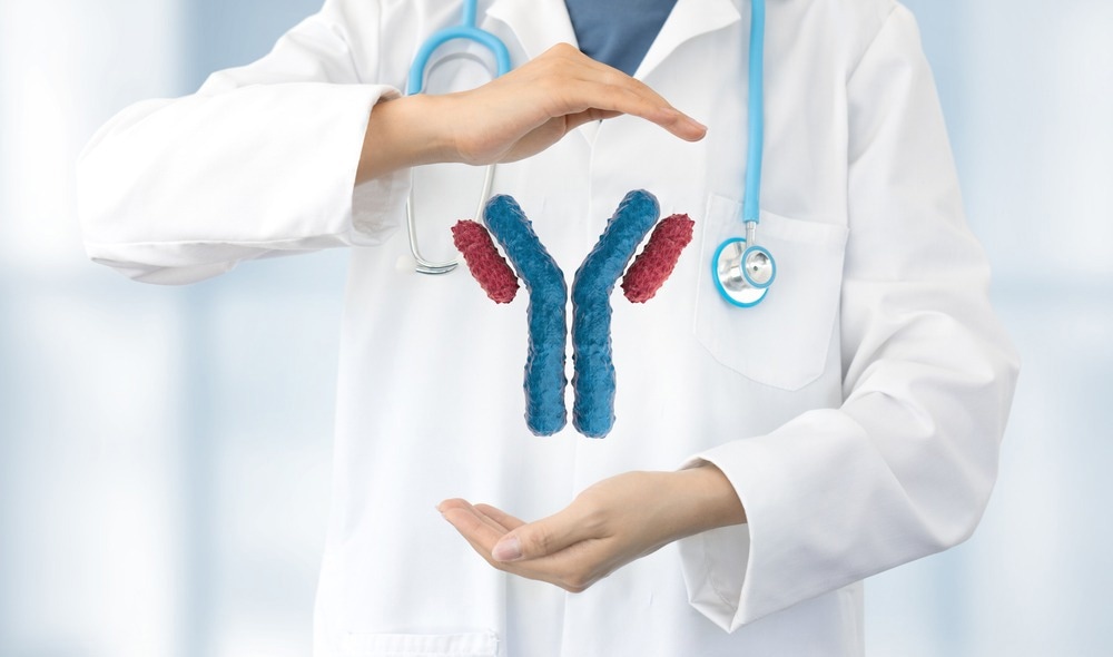
In a recent Scientific Reports journal paper, researchers isolated 7G7 and NIBIC-71 from peripheral blood mononuclear cells (PBMCs) derived from coronavirus disease 2019 (COVID-19) convalescent individuals. These monoclonal antibodies (mAbs) were found to neutralize the severe acute respiratory syndrome coronavirus 2 (SARS-CoV-2) Delta and Omicron variants.

Study: Human antibody recognition and neutralization mode on the NTD and RBD domains of SARS-CoV-2 spike protein. Image Credit: paulista / Shutterstock.com
Background
The continual emergence of more immune-evasive and transmissible SARS-CoV-2 variants of concern (VOCs), such as Delta and Omicron, has threatened the efficacy of COVID-19 vaccines and anti-SARS-CoV-2 therapeutic agents. Neutralizing mAbs targeting the receptor-binding domain (RBD) of the SARS-CoV-2 spike (S) glycoprotein, which comprises S1 and S2 subunits, have been developed to curtail SARS-CoV-2 transmission; however, optimal target regions on SARS-CoV-2 S need to be identified for the effective neutralization of SARS-CoV-2 VOCs.
Previous studies have reported that inhibiting SARS-CoV-2 S-angiotensin-converting enzyme 2 (ACE2) interactions can suppress SARS-CoV-2 infections; however, data on COVID-19 suppression by suppressing S1/S2 cleavage are lacking.
About the study
In the present study, researchers isolated two mAbs, 7G7 and NIBIC-71, that neutralized SARS-CoV-2 VOCs through different neutralization mechanisms.
PBMCs were extracted from two COVID-19 convalescents. Anti-S antibody-producing lymphoblastoid cell lines (LCLs) were prepared by infecting B lymphocytes with Epstein-Barr virus (EBV).
Enzyme-linked immunosorbent assays (ELISA) were performed to identify anti-S LCLs. In addition, the neutralizing activity of the mAbs was assessed using cytopathic effect (CPE) inhibition assays.
In addition, antibody amino acid sequencing and B-cell receptor (BCR) amplicon sequencing analyses were performed by liquid chromatography/mass spectrometry (LC-MS) analysis to detect S-targeted mAb sequences in the LCL supernatants. Subsequently, the team integrated sequence data and assessed DNA sequences of 7G7 and NIBIC-71 based on which somatic hypermutation (SHM) rates were calculated.
Recombinant mAbs NIBIC-71, 7G7/λ, and 7G7/κ were prepared to characterize 7G7 and NIBIC‑71. In addition, s-ACE2 interactions were assessed using ELISA.
In addition, the binding affinities of the two mAbs with SARS-CoV-2 S were evaluated by surface plasmon resonance (SPR) analysis. Several truncated S proteins were also prepared to identify SARS-CoV-2 S regions identified as epitopes by the two mAbs using ELISA. Finally, cryo-electron microscopy (cryo-EM) was also performed to assess the mAbs’ binding modes to SARS-CoV-2 S.
The inhibition of SARS-CoV-2 host cell invasion by the two mAbs was evaluated in vitro by luciferase activity-based neutralizing assays. S-ACE2 binding inhibition and the suppression of S1/S2 cleavage by the two mAbs were also assessed.
Lab Diagnostics & Automation eBook

The neutralizing potency of 7G7/κ and NIBIC-71 was also evaluated in vivo by infecting Syrian hamsters with SARS-CoV-2 wild-type (WT) strain intranasally after injecting the two mAbs and an additional control mAb intravenously.
Pulmonary tissues of the infected hamsters were obtained and subjected to histological analysis. These tissues were also used to determine the pulmonary viral load through CPE and reverse transcription-polymerase chain reaction (RT-PCR) assays.
SARS-CoV-2 VOC neutralization by the two mAbs was evaluated by infecting VeroE6-transmembrane protease serine 2 (TMPRSS2) cells with SARS-CoV-2 WT, Delta, and Omicron BA.1.
Study findings
In total, 28 S-specific LCL supernatants were detected using ELISA, two of which inhibited SARS-CoV-2-associated CPE effects. Both 7G7 and NIBIC-71 mAbs effectively neutralized SARS-CoV-2 WT and Delta, but not Omicron.
NIBIC-71 was RBD-specific, whereas 7G7/κ targeted the N-terminal domain (NTD) of S1. NIBIC-71 inhibited S-ACE2 binding, whereas 7G7/κ did not, and 7G7/κ suppressed S1/S2 disintegration, but NIBIC-71 did not.
The NIBIC-71 mAb was derived from immunoglobulin G1 (IgG1)/Igκ with Ig Kappa variable 1-9 (IGKV1-9) and Ig heavy variable 3-53 (IGHV3-53) genes. The 7G7 (7G7/λ) mAb was derived from IgG1/Igλ2 with Ig lambda variable 3-25 (IGLV3-25) and IGHV3-33 genes.
The 7G7/κ, NIBIC-71, and 7G7/λ mAbs had similar half-maximal effective concentrations (EC50) values. A dose-dependent response against SARS-CoV-2 S was observed, thus indicating that the mAbs were S-specific. The binding affinity of 7G7/κ (IgG1/Igκ) for SARS-CoV-2 S was higher as compared to 7G7/λ (IgG1/Igλ2).
Cryo-EM findings of 7G7/κ and NIBIC-71 complexes at 2.6 Å and 3.0 Å resolution, respectively, showed that two NIBIC-71 fragment antigen-binding regions (Fabs) interacted with two up conformation-S RBDs, whereas all three Fabs of 7G7/κ were bound to the three NTDs of SARS-CoV-2 S. Additionally, NIBIC-71 had a greater half-maximal inhibitory concentration (IC50) value as compared to 7G7/κ.
In vivo findings showed no statistically significant differences between the mAbs concerning rectal temperature and bodyweight alterations in the hamsters. Both mAbs lowered SARS-CoV-2 viral loads significantly more than the control.
The SARS-CoV-2 nucleocapsid protein was detectable in control mAb-infected hamster lungs but not in the lungs of NIBIC-71- or 7G7/κ-treated hamsters. Histological analysis showed that NIBIC-71 and 7G7/κ reduced COVID-19-induced inflammation.
The mode of binding for NIBIC-71 was unaffected by Delta VOC mutations T478K and L452R. Furthermore, multiple mutational sites in the Omicron VOC including G446S, K417N, Q493R, S477N, Q498R, G496S, Y505H, and N501Y overlapped with NIBIC-71’s epitope.
The T19R mutation in Delta may have affected 7G7/κ binding. Additionally, the deletion of residues 142 to 144, as well as the Y145D amino acid substitution in the Omicron VOC, likely altered the 7G7/κ surface at the point of complementarity-determining region (CDR) contact.
Overall, the study findings demonstrate the likelihood of two novel neutralizing mAbs targeting S1/S2 cleavage being cross-reactive against SARS-CoV-2 VOCs.
- Otsubo, R., Minamitani, T., Kobiyama, K. et al. (2022). Human antibody recognition and neutralization mode on the NTD and RBD domains of SARS-CoV-2 spike protein. Scientific Reports. doi:10.1038/s41598-022-24730-4
Posted in: Medical Science News | Medical Research News | Disease/Infection News
Tags: ACE2, Amino Acid, Angiotensin, Angiotensin-Converting Enzyme 2, Antibodies, Antibody, Antigen, binding affinity, Blood, Cell, Chromatography, Coronavirus, Coronavirus Disease COVID-19, covid-19, DNA, Efficacy, Electron, Electron Microscopy, ELISA, Enzyme, Epstein-Barr Virus, Genes, Glycoprotein, Immunoglobulin, in vivo, Inflammation, Liquid Chromatography, Luciferase, Lungs, Mass Spectrometry, Microscopy, Mutation, Omicron, Polymerase, Polymerase Chain Reaction, Protein, Receptor, Respiratory, SARS, SARS-CoV-2, Serine, Severe Acute Respiratory, Severe Acute Respiratory Syndrome, Spectrometry, Syndrome, Transcription, Virus

Written by
Pooja Toshniwal Paharia
Dr. based clinical-radiological diagnosis and management of oral lesions and conditions and associated maxillofacial disorders.
Source: Read Full Article
