Blood carries vital oxygen through our circulation system to muscles and organs. Acoustic tools can create small bubbles in our blood, capable of changing in response to oxygen and signifying oxygen levels.
During the 181st Meeting of the Acoustical Society of America, which will be held Nov. 29 to Dec. 3, Shashank Sirsi, from the University of Texas at Dallas, will discuss how circulating microbubbles can be used to measure oxygen levels. The talk, “Hemoglobin Microbubbles for In Vivo Blood Oxygen Level Dependent Imaging: Boldly Moving Beyond MRI,” will take place Monday, Nov. 29.
Microbubbles are smaller than one hundredth of a millimeter in diameter and can be made by emulsifying lipids or proteins with a gas. The gas filling of microbubbles causes them to oscillate and vibrate when ultrasound is applied, scattering energy and generating an acoustic response that can be detected by a clinical ultrasound scanner. They are routinely used in medical imaging to provide greater contrast in tissue.
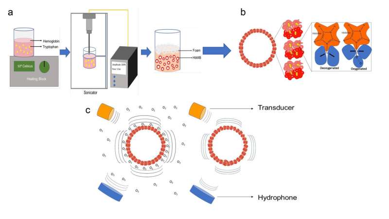
Hemoglobin, the protein that gives red blood cells their signature color, will form a stable shell around microbubbles. It then continues to carry out its typical role of binding and releasing oxygen in blood.
Sirsi and his team developed microbubbles to acoustically detect blood oxygen levels, since the microbubble shells are altered by structural hemoglobin changes in response to oxygen. The hemoglobin shell is continually responsive to oxygen after surrounding the bubble and has been optimized to perform in living organisms’ circulation.
“When oxygen binds to hemoglobin, there are structural changes in the protein that change the mechanical properties,” said Sirsi. “The mechanical properties of the shell dictate the acoustic response of a bubble, so our hypothesis was that different acoustic responses would be seen as the shell gets stiffer or more elastic.”
Source: Read Full Article
