Metastasis occurs when cancer cells acquire the ability to spread and form new tumors in different places in the body, usually by traveling within blood or lymph vessels. Since metastasis is a hallmark of advanced cancer and severely complicates treatment, its early diagnosis is essential. One way to do this is by looking for circulating tumor cells (CTCs) in blood samples.
However, CTCs can be very rare, and they might be completely absent in small blood samples despite being present in a patient’s bloodstream. To address this problem, researchers have developed a technique called diffuse in-vivo flow cytometry (DiFC). It involves labeling CTCs with a fluorescent agent, shining a laser directly onto an artery, and capturing the emitted fluorescent signals using a detector to count the number of CTCs.
While promising, DiFC measurements can be severely affected by background noise originating mainly from the inherent fluorescence of the surrounding tissue called autofluorescence (AF).
A collaborative research team from Tufts University and Northeastern University in Massachusetts, U.S., has been actively trying to address this issue and take the DiFC method developed by the Northeastern group to a new level. In their latest study, published in Journal of Biomedical Optics (JBO), the researchers tested the capabilities of a new method developed by the Tufts group called the dual-ratio (DR) approach in minimizing noise in DiFC and thereby extending its penetration range.
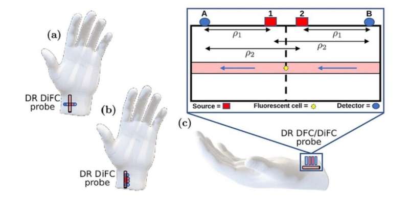
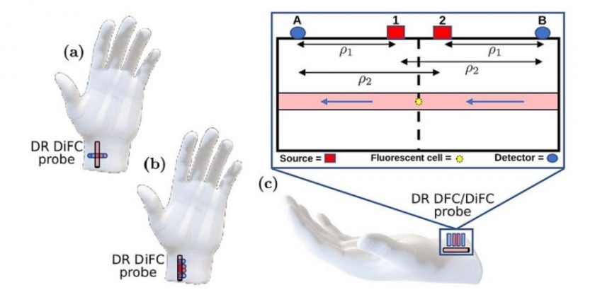
The DR approach was originally conceived for spectroscopy techniques and has now been adopted for DiFC. The resulting DR DiFC system employs two laser sources and two detectors arranged carefully in space. In theory, the noise can be canceled out by combining the signals of the two detectors, and the AF contributions of the surface of the measured medium, say, skin, can also be minimized using the DR approach. However, the conditions under which DR DiFC truly offers an advantage over standard DiFC remain unclear.
The researchers addressed this knowledge gap in three ways. They ran Monte-Carlo simulations using various noise and AF parameters, as well as for different source–detector configurations. They also conducted DR DiFC experiments using an artificial tissue-mimicking flow phantom with cell-mimicking fluorescent microspheres. Lastly, the team measured the AF of the skin and the underlying muscle of mice to gain insight into the variation of noise with tissue type and depth.
The experiments revealed that DR DiFC was superior to standard DiFC if the fraction of noise not canceled by DR was under 10 percent and if the contributions to AF were surface-weighted—higher near the surface rather than being evenly distributed in the target volume.
However, as the experiments in mice suggested, AF is typically much higher in skin than in the underlying muscle, implying that DR DiFC may offer an advantage over standard DiFC in most cases. Notably, if AF was higher near the surface rather than being homogeneous, DR DiFC had a significantly higher penetration range than standard DiFC.
Overall, the findings of this study will be pivotal in the development of DR DiFC as an emerging technique to noninvasively detect fluorescent molecules in the bloodstream. This method will enable doctors to quickly detect cancer cells in the blood of patients without having to draw samples, making the diagnosis of metastasis simpler and more accurate. It could be extended to other cell types or even systemic molecules of interest in the future.
More information:
Giles Blaney et al, Dual-ratio approach for detection of point fluorophores in biological tissue, Journal of Biomedical Optics (2023). DOI: 10.1117/1.JBO.28.7.077001
Journal information:
Journal of Biomedical Optics
Source: Read Full Article
