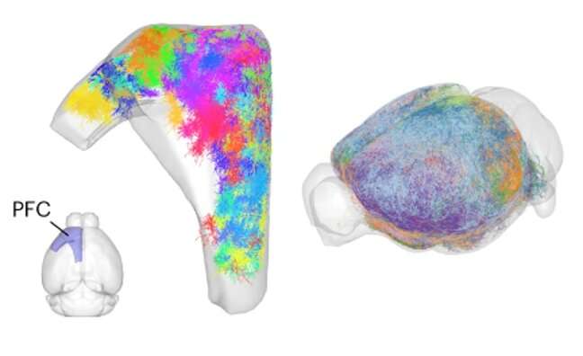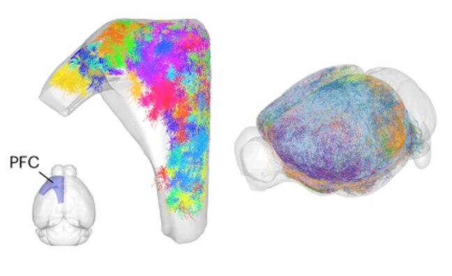
The prefrontal cortex (PFC) is a crucial part of the mammalian brain, known to be involved in decision-making, planning, complex social behavior and integrating the activity of other brain regions. While countless neuroscience studies have investigated the function and structure of this key brain region, much about the organization of neurons within it is yet to be uncovered.
Researchers at the Chinese Academy of Sciences have recently been conducting research aimed at better understanding how axons (i.e., regions of neurons that generate and transmit impulses) and dendrites (i.e., branches extending from neurons that receive impulses from other neurons) are organized in the PFC. In a recent paper published in Nature Neuroscience, they mapped thousands of dendrites and axons in the mouse PFC using an innovative tool they developed.
“Our previous paper reconstructed the complete axons of more than 6,000 neurons in mouse PFC,” Jun Yan, one of the researchers who carried out the study, told Medical Xpress. “The same data also contain the dendrites of these neurons. As the dendrite morphology plays an important role in defining the way neurons receive information, we thought it is natural as a next step to reconstruct the dendrites of these neurons and investigate the relationship between axons and dendrites.”
To conduct their new analyses, the researchers used data that they collected as part of a previous study, using a tool that they developed called “fast neurite tracer” (FNT). This is essentially a software tool that can trace tera-bytes of images via a three-dimensional (3D) graphical interface.
“We analyzed the same data in which we previously traced axons, this time selecting about 2,000 neurons with the best image quality of dendrites for complete reconstruction,” Yan explained. “We then classified these dendrites based on their morphology and analyzed their correspondence with the classification based on axon projections for the same neurons.”
Through their analyses, Yan and his colleagues were able to identify 1,515 pyramidal projection neurons and 405 atypical pyramidal projection neurons or spiny stellate neurons with unconventional axon projection patterns. In addition, they gathered new insights on the organization of projection neurons in the mouse PFC, showing that these neurons could communicate with neurons in their same column and in other columns, as well as in other brain regions, allowing neural information to flow in a specific manner across PFC.
“We identified both shared and divergent features between dendrite morphology and axon projection,” Yan said. “Our study has two key implications. Firstly, it shows that a more comprehensive neuron classification should consider dendrites and axons together. Secondly, the availability of both dendrites and axons allowed us to better reconstruct a neural network.”
The recent findings gathered by this team of researchers offer a detailed reconstruction of axons and dendrites in the mouse PFC, which could inform future neuroscience research and theoretical predictions. This could in turn improve the present understanding of the PFC of all mammals, including humans, while also potentially informing the development of new sophisticated brain-inspired computational tools.
“The next step of this study would be to validate the neural network we reconstructed from dendrites and axons and learn the functions of the neural network,” Yan added. “For example, can this PFC neural network teach us about the new network design for artificial intelligence?”
More information:
Le Gao et al, Single-neuron analysis of dendrites and axons reveals the network organization in mouse prefrontal cortex, Nature Neuroscience (2023). DOI: 10.1038/s41593-023-01339-y
Journal information:
Nature Neuroscience
Source: Read Full Article
