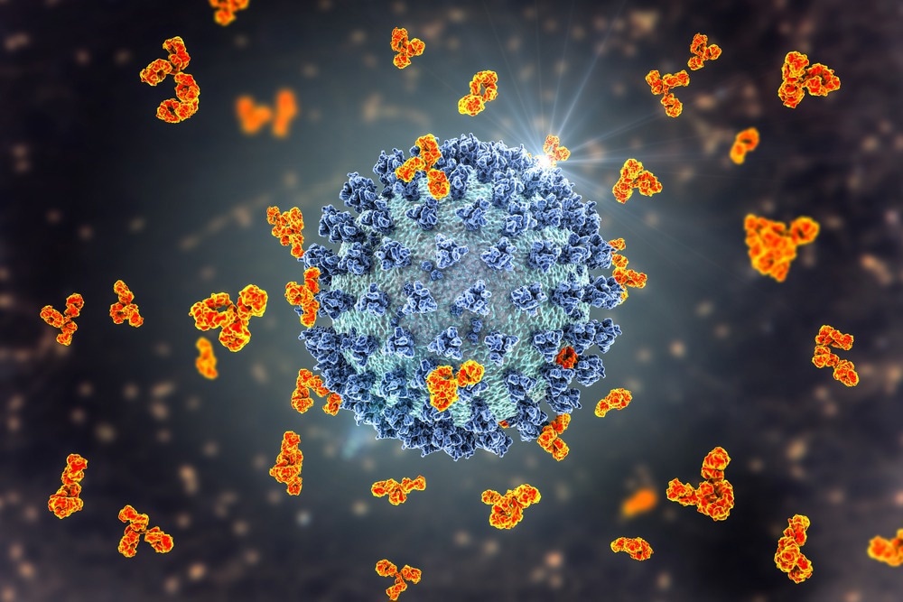
In a recent study published in PLoS ONE, researchers developed humanized nanobodies that neutralize severe acute respiratory syndrome coronavirus 2 (SARS-CoV-2).

Background
The coronavirus disease 2019 (COVID-19) pandemic has caused a global health emergency at an unprecedented scale. Consequently, the scientific community responded by rapidly developing SARS-CoV-2 vaccines. Although SARS-CoV-2 vaccination has been effective against severe COVID-19 and associated hospitalization, there is potential for the emergence of escape variants.
Therefore, there is a pressing need for affordable, high-throughput pipelines to explore effective anti-SARS-CoV-2 therapeutics. Nanobodies are unique heavy-chain only antibodies in camelids and some shark species. As anti-viral biologics, nanobodies offer several advantages such as long shelf life, convenient mass production in prokaryotic systems, and higher tissue permeability.
The study and findings
In the present study, researchers described an efficient, rapid strategy to design and develop SARS-CoV-2-neutralizing humanized nanobodies. First, phage-displayed nanobody libraries were prepared, starting from a caplacizumab-derived backbone, to establish a platform for the rapid discovery of nanobodies.
One round of phage panning was performed against the SARS-CoV-2 spike S1 domain, with two additional rounds against the receptor-binding domain (RBD). The authors identified 13 nanobodies as a result of panning. Nanobodies were synthesized and purified by Ni-NTA affinity chromatography and size-exclusion chromatography.
Global fit modeling revealed that binding affinities towards RBD-mFc were 0.9 nM for RBD-1-1E nanobody and 9.4 nM for RBD-1-2G. RBD-1-2G had an affinity of 6.9 nM to the S1-Fc analyte, a 26.6% improvement over RBD-mFc. Further, AlphaLISA results indicated that RBD-1-2G nanobody was a potential therapeutic candidate.
Next, bivalent and trivalent modalities of RBD-1-2G were constructed for further evaluation or improved neutralization. The bivalent version (RBD-1-2G-Fc) showed improved affinity (1.9 nM), and the trivalent version (RBD-1-2G-Tri) showed further improvement with an affinity of 0.1 nM for RBD. The authors next investigated whether the improved affinity corresponded to increased therapeutic potential. To this end, an endocytosis assay was performed using SARS-CoV-2 RBD quantum dots.
RBD-1-2G exhibited the highest potency with a half-maximal inhibitory concentration (IC50) of 390 nM. The IC50 was further reduced to 14 nM with the bivalent version (RBD-1-2G-Fc). RBD-1-2G neutralized SARS-CoV-2 pseudoviruses with an IC50 of 490 nM, and significant improvements were evident with RBD-1-2G-Fc (IC50 – 88 nM) and RBD-1-2G-Tri (IC50 – 4.1 nM).
In a live virus neutralization screen, the trivalent (IC50 – 182 nM) and bivalent (IC50 – 255 nM) constructs were more potent than RBD-1-2G (IC50 – 1211 nM). A human membrane proteome array was performed to assess the reactivity and specificity of RBD-1-2G. This screening confirmed that RBD-1-2G showed high specificity to SARS-CoV-2 spike protein with minimal binding to membrane proteins.
Notably, the nanobody was bound to mitochondrial elongation factor 1 (MIEF1) in the initial screening. This assay showed that RBD-1-2G could bind to potential off-targets. Cryo-electron microscopy was performed to obtain density maps of soluble spike ectodomain complexed with four nanobodies.
The accessible epitopes were classified into groups 1 and 2. Group 1 epitope comprised the binding region of RBD-1-2G, RBD-1-1E, and RBD-2-1F nanobodies at the distal end of ‘up’ RBD and in an area overlapping the receptor-binding motif (RBM).
RBD-1-1G and RBD-1-3H bound epitopes (group 2) were exposed on the face of erect RBD in a region that did not overlap with RBD. The atomic fitting model of the spike revealed that group 1-binding nanobodies could overlap with the angiotensin-converting enzyme 2 (ACE2) binding site, whereas group 2 binding nanobodies do not inhibit ACE2 binding.
Furthermore, RBD-1-2G-Tri inhibited SARS-CoV-2 pseudo-typed particles better than wild-type particles. RBD-1-2G and its bivalent version had similar IC50 values against the Alpha variant. They designed a three-dimensional (3D) tissue model to better replicate the complex mammalian respiratory tract tissues.
The human airway air-liquid interface (ALI) tissues were infected with 5000 plaque-forming units (PFU) of SARS-CoV-2 WA1 strain or Alpha variant in the presence of RBD-1-2G and its bi- and trivalent constructs. RNA levels of the viral nucleocapsid gene were determined by quantitative reverse transcription polymerase chain reaction (qRT-PCR).
The delta cycle threshold (dCt) value of WA1-infected cells was four cycles in the no nanobody treatment group; this increased to 20.4 (RBD-1-2G), 22.8 (bivalent), and 17.2 (trivalent) cycles after treatment. Similar trends were noted for Alpha-infected cells.
The average dCt of Alpha-infected cells in the no-treatment group was 2.2 cycles, meaning that these cells had nearly 3.5 times more viral RNA than WA1-infected cells. Overall, RBD-1-2G-Fc had better activity than RBD-1-2G-Tri in the ALI model, indicating that the bivalent construct might be favorable for clinical transition.
Conclusions
In summary, the study isolated and characterized SARS-CoV-2-neutralizing nanobodies from a pool of humanized phage libraries. The nanobodies bound to the spike RBD with exceptional affinity, neutralized pseudo-typed particles and authentic SARS-CoV-2. RBD-1-2G exhibited the best potency among the nanobodies and improved neutralization when constructed into bivalent and trivalent modalities.
The nanobody bound to an epitope on the top of RBD, overlapping with the ACE2 binding site. RBD-1-2G’s potency was attributed to its high binding affinity and the ability to bind to RBD in different conformations. Overall, the increased activity profile, with binding in multiple RBD conformations, suggested that RBD-1-2G could be an ideal prototype for new candidates capable of adapting to numerous SARS-CoV-2 variants.
- Fu, Y. et al. (2022) "A humanized nanobody phage display library yields potent binders of SARS CoV-2 spike", PLOS ONE, 17(8), p. e0272364. doi: 10.1371/journal.pone.0272364. https://journals.plos.org/plosone/article?id=10.1371/journal.pone.0272364
Posted in: Medical Science News | Medical Research News | Disease/Infection News
Tags: ACE2, Analyte, Angiotensin, Angiotensin-Converting Enzyme 2, Antibodies, Assay, binding affinity, Chromatography, Coronavirus, Coronavirus Disease COVID-19, covid-19, Electron, Electron Microscopy, Enzyme, Gene, Global Health, Membrane, Microscopy, Nanobodies, Pandemic, Polymerase, Polymerase Chain Reaction, Protein, Proteome, Quantum Dots, Receptor, Respiratory, RNA, SARS, SARS-CoV-2, Severe Acute Respiratory, Severe Acute Respiratory Syndrome, Spike Protein, Syndrome, Therapeutics, Transcription, Virus

Written by
Tarun Sai Lomte
Tarun is a writer based in Hyderabad, India. He has a Master’s degree in Biotechnology from the University of Hyderabad and is enthusiastic about scientific research. He enjoys reading research papers and literature reviews and is passionate about writing.
Source: Read Full Article
